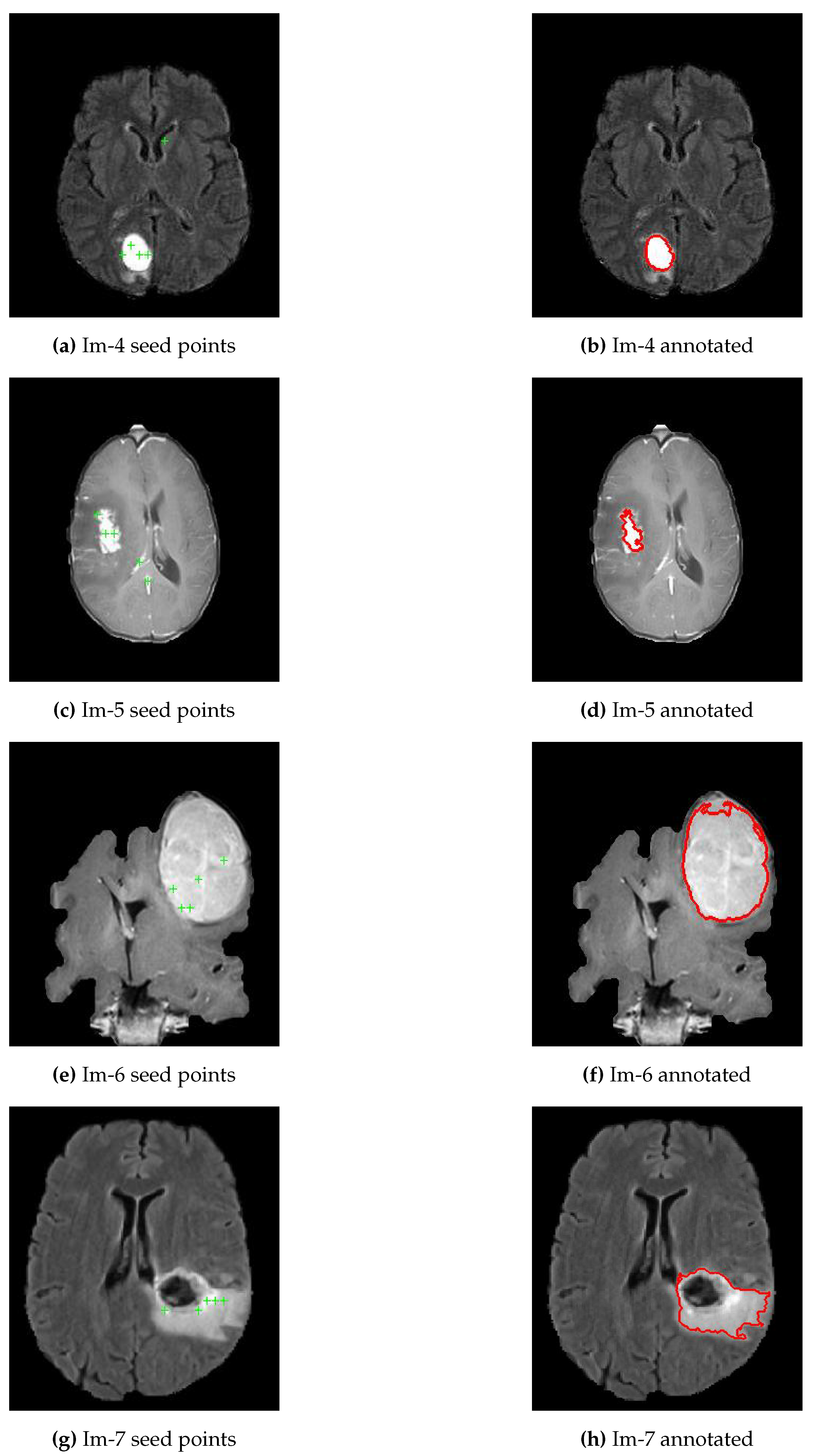
Lesion region Then, histogram thresholding is acquired to automate the seeds selection for region growing process The region is iteratively grown by comparing all unallocated neighboring pixels to the seeds The difference between pixel's intensity value and the region's mean is used as the similarity measureSpontaneous Activity Within Seed Region!This hypothesis was evaluated using diffusion Magnetic Resonance Imaging (dMRI) tractography in two successive samples of right and lefthanded subjects randomly selected from the Human Connectome Project (HCP) database The dorsoposterior parietal hand control region (DPPC hand) was used as the seed region of interest (ROI) for the tractography

European Ultrahigh Field Imaging Network For Neurodegenerative Diseases Eufind Duzel 19 Alzheimer S Amp Dementia Diagnosis Assessment Amp Disease Monitoring Wiley Online Library
Seed region mri
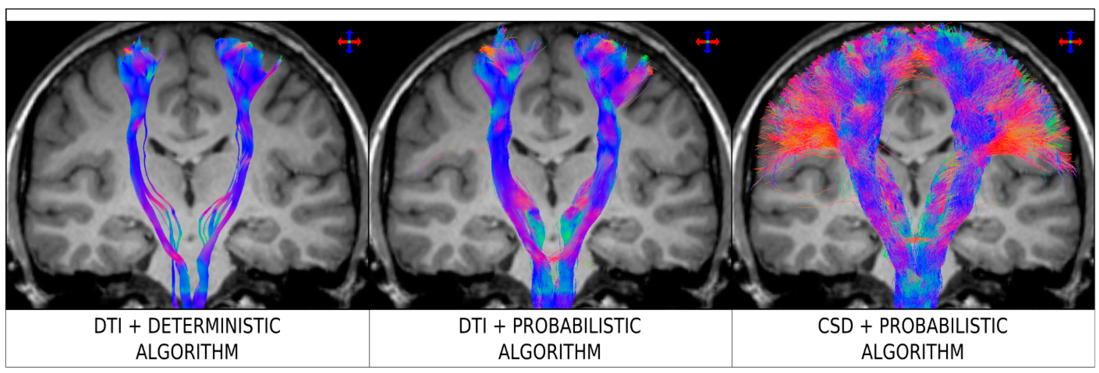
Seed region mri-Diffusion MRI fiber tracking uses local fiber directions measured at each voxel to track the trajectory of a white matter pathway If we set region A as the seed region and region B as the ending region, does this find tracks ending in both regions? Seedbased functional connectivity, also called ROIbased functional connectivity, finds regions correlated with the activity in a seed region In seedbased analysis, the crosscorrelation is computed between the timeseries of the seed and the rest of the brain (telling us where the traffic is communicating between selected cities) (Fig 3


Post Mortem Mapping Of Connectional Anatomy For The Validation Of Diffusion Mri Biorxiv
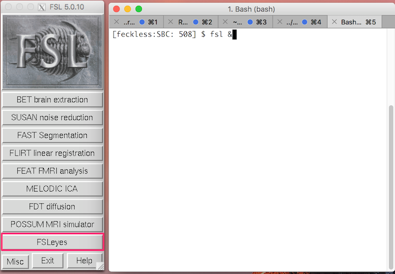
The seed region will be the Posterior Cingulate Gyrus You will identify the seed region using the HarvardOxford Cortical Atlas To start FSL, type the following in the terminal fsl & NB The & symbol allows you to type commands in this same terminal, instead of having to open a second terminal Click FSLeyes to open the viewerFcMRI signals while regions that become negative when a seed regions activates tend to be negatively correlated with the seed region (Fox et al, 06;*STEP 3 use mri_annotation2label to create labels from the new annotation This will create a label for each parcellation (including labels for regions outside of your desired seed region) The labels will be deposited in the subject's recondir/label directory Sample command mri_annotation2label subject \
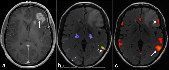
Instructions Create two separate axial seed regions (at approx axial slice 64), one for each side Create one ROA region and draw a sagittal ROA slice at the midline In the region list, check only the left seed region, then run fiber tracking Based on this output, ROA placement will be clearer on a coronal slice superior to the seed regionSegmentation Region Growing where $\mu$ is the mean intensity of the seed points, We first load a T1 MRI brain scan and select our seed point(s) If you are unfamiliar with the anatomy you can use the preselected seed point specified below, just uncomment the line PURPOSE Restingstate functional MRI (rsFMRI) has shown potential for presurgical mapping of eloquent cortex when a patient's performance on taskbased FMRI is compromised The seedbased analysis is a practical approach for detecting rsFMRI functional networks;
The region growing method can work efficiently in medical image segmentation if one can guarantee optimal initial seed point and threshold criterion used to stop growing outside a region In seeded region growing method (SRG), seed selection is crucial, often done by hand in medical image processing , 21Greicius et al, 03) Because "seeds" can be placed in any cortical or subcortical location, development of methods for analyzing these correlation3 Seed region growing The basis of the method is to segment an image of N pixels into regions with respect to a set of seeds 26 using only the initial seed pixels The initial seed pixel is selected from a pixel with mask 3X3 These seeds are grown by merging neighboring pixels whose properties are most similar to the


Resting State Fmri Wikipedia


High Connectivity Between Reduced Cortical Thickness And Disrupted White Matter Tracts In Long Standing Type 1 Diabetes Diabetes
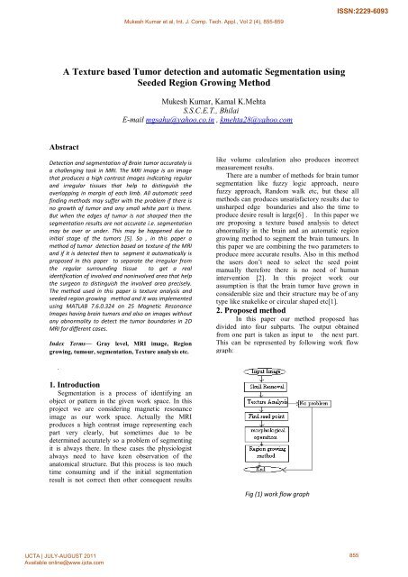
The seed point can be manually selected by an operator or automatically initialised with a seed finding algorithm Then, region growing examines all neighboring pixels/voxels and if their intensities are similar enough (satisfying a predefined uniformity or homogeneity criterion), they are added to the growing region "Unsupervised MRI Cerebral responses to putative pheromones and objects of sexual attraction were recently found to differ between homo and heterosexual subjects Although this observation may merely mirror perceptional differences, it raises the intriguing question as to whether certain sexually dimorphic features in the brain may differ between individuals of the same sex butThe seed point pixels are analyzed by the predetermined region growing formula When all the neighboring pixels are included into the seed pixels domain, at that point the region is said to be grown and region growing stops Fig6 Tumor detected after region growing algorithm TPFPTNFN STEP 6 Edge detection of tumor


A Window Into The Brain Advances In Psychiatric Fmri


Resting State Seed Based Analysis An Alternative To Task Based Language Fmri And Its Laterality Index American Journal Of Neuroradiology
In the Z direction We thus determine the number of seeds in each region by analyzing the region volume statistics Once each region s seed count is determined, the region is divided into equal subregions along the Z axis, where nally, the centroids of each subregion determine the seed coordinates 23 Experiment We built two MRI compatible Actually my project is brain tumor segmentation in MRI images I want to segment the brain MRI images using region growing technique How can I find a better seed point that detects the brain tumor efficientlySample images are attached 0 Comments Show HideSeeded Region Growing is an integrated method brought up by Adams and Bischof 413, in which few initial seeds are generated, and more similar neighboring regions are then combined to achieve region growing 1421 In addition, the method of unsupervised vector seeded region growing suitable for medical multispectral images was established


Automatic Region Based Brain Classification Of Mri T1 Data


Figure 3 From A Window Into The Brain Advances In Psychiatric Fmri Semantic Scholar
2) Based upon the selected seed point, whole image get split into foreground and background region 3) Foreground region is Figure 2 MRI of human body then enhanced by equalizing histogram adaptively and then Results background region is added to the enhanced foregroundSeeded Region Growing (SRG) algorithm, originally proposed by Adams and Bischof 6, is a fast, robust, parameterfree method for segmenting intensity images given initial seed locations for each region In SRG, individual pixels that satisfy some neighborhood constraint are merged if their attributes, such as intensity or texture,$ 40 30 10 0 10 30 40 0 5 10 15 25 30 35 40 45 50 55 60 65 70 75 80 85 90 Biswal$etal,$MRM,$1995$ How$
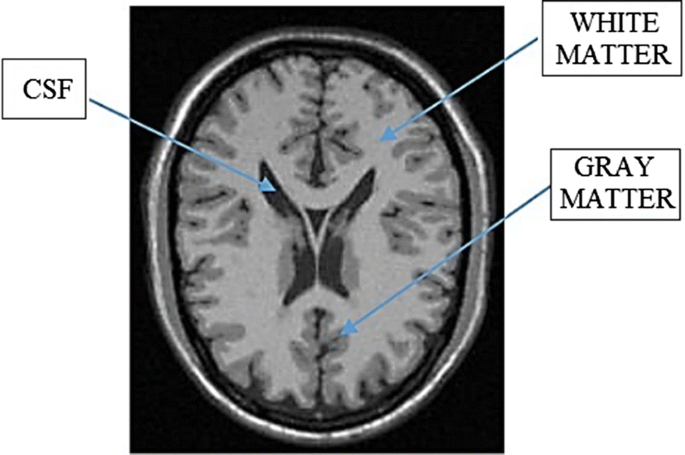


Automatic Seeded Region Growing Asrg Using Genetic Algorithm For Brain Mri Segmentation Springerlink


A Cade System For Gliomas In Brain Mri Using Convolutional Neural Networks Deepai
In this paper, an automatic computeraided detection system for breast magnetic resonance imaging (MRI) tumour segmentation will be presented The study is focused on tumour segmentation using the modified automatic seeded region growing algorithmYes, it is safe to have a colonoscopy, however, we recommend waiting 6 months after the seed implant before having a colonoscopy• Results are not symmetric between "seed" and "target" regions • Sensitive to areas of high local uncertainty in orientation (eg, pathaway crossings), errors propagate from those areas • Best suited for exploratory tractography studies • All connections from a seed region, not constrained to a specific target region
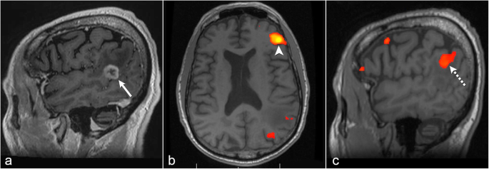


The Role Of Resting State Functional Mri For Clinical Preoperative Language Mapping Cancer Imaging Full Text


Maps Of Seed Based Resting State Fmri Functional Connectivities The Download Scientific Diagram
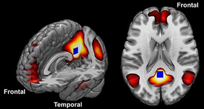
Abstract In investigations of the brain's resting state using functional magnetic resonance imaging (fMRI), a seedbased approach is commonly used to identify brain regions that are functionally connected The seed is typically identified based on anatomical landmarks, coordinates, or the location of brain activity during a separate taskThe map represents a restingstate functional connectivity analysis performed on 1,000 human subjects, with the seed placed at the currently selected location Thus, it displays brain regions that are coactivated across the restingstate fMRI time series with the seedThe present study hypothesizes that structural connectivity from key seed regions may induce effects on their connected targets, which are reflected in gene expression at those targeted regions To test this hypothesis, analyses were performed on data from two brains from the Allen Human Brain Atlas, for which both gene expression and DWMRI
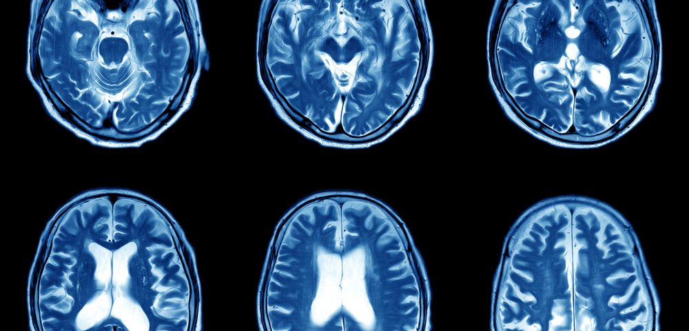


Early Mris May Predict 9 Year Outcomes In Ms Children Study Says
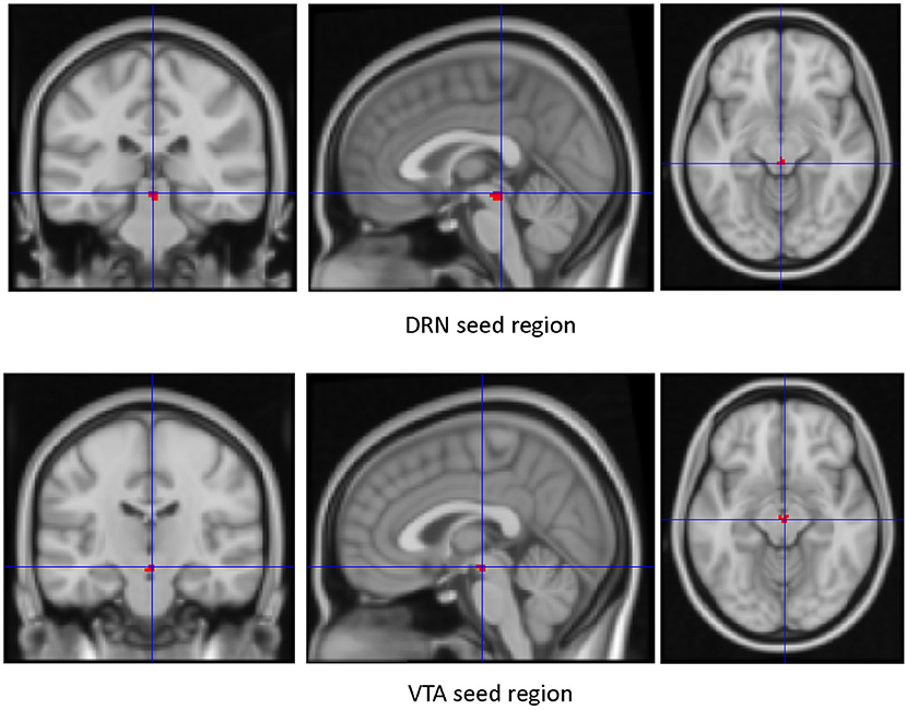


Frontiers Resting State Functional Connectivity Of Dorsal Raphe Nucleus And Ventral Tegmental Area In Medication Free Young Adults With Major Depression Psychiatry
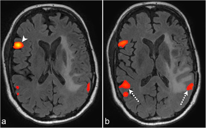
(A, B) The restingstate functional magnetic resonance imaging using a seed region on Broca's area shows Wernicke's area displaced posteriorly and superiorly by the tumor (arrow in B) (C, D) The tumor was resected under iMRI control with preservation of the eloquent cortex iMRI, intraoperative magnetic resonance imagingHowever, seed localization remains challenging for presurgical language mappingRisks of Spinal MRI MRI scans are considered safer than CT scans or Xrays However, there are risks MRIs use a magnet that creates a strong magnetic field
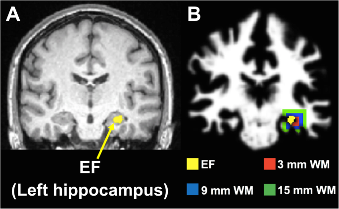


18 F Fdg Pet Guided Diffusion Tractography Reveals White Matter Abnormalities Around The Epileptic Focus In Medically Refractory Epilepsy Implications For Epilepsy Surgical Evaluation European Journal Of Hybrid Imaging Full Text


1
We applied novel fetal restingstate functional MRI to measure brain function in 32 human fetuses in utero and found that systemslevel neural functional connectivity wasSeed selection process Region based segmentation of the medical images is widely used in various clinical applications such as bone and SemiAutomatic Seeded Region Growing for Object Extracted in MRI International Journal of Scientific & Engineering Research Volume 7, Issue 2, February16MRI using taskbased or stimulusdriven paradigms has been time course of the seed region and that of all other areas in the brain, the authors found that the left somatosensory cortex was RSNs Compared with seedbased methods, ICA has the advantage of requiring few a priori assumptions but does compel the


Success Of 125i Seed Treatment In Vulvar Squamous Cell Carcinoma With Ott


European Ultrahigh Field Imaging Network For Neurodegenerative Diseases Eufind Duzel 19 Alzheimer S Amp Dementia Diagnosis Assessment Amp Disease Monitoring Wiley Online Library
3 Probabilistic fiber trackingThe method of seedbased functional connectivity studies regions correlated with the activity in a seed region In seedbased analysis, the crosscorrelation is identified between the timeseries of the seed and the rest of the brain 9 Zang et al found that the activation of bilateral cerebellum was decreased in ADHD group 10 Therefore, this Global Root Vegetable Seeds Market report has covered and analyzed the potential of Worldwide market Industry and provides statistics and information on market dynamics, market analysis, growth factors, key challenges, major drivers & restraints, opportunities and forecastThis report presents a comprehensive overview, market shares, and growth opportunities of market


Post Mortem Mapping Of Connectional Anatomy For The Validation Of Diffusion Mri Biorxiv


Seeds Regions Of Interest Used In The Study A Default Mode Network Download Scientific Diagram
1 day ago GMO Seed Market – Top Leading Countries WMR mohit 2 Exclusive GMO Seed Market research report is a significant source of keen information for business specialists It furnishes the business outline with development investigation and historical and futuristic cost analysis, revenue, demand, and supply information ThisFigure 1 MRI of a Brain Tumor Patient Figure 2 Segmented Tumor and Posterior Orbital Fat Regions of the MRI Image in Figure 1 23 Intensity Space Map (ISM) Region growing segmentation techniques have been used to extract a connected region of similar pixels from an image Mancas et al 7 proposed a region growing segmentation Instructions Create two separate axial seed regions (at approx axial slice 99), one for each side Create one ROA region and draw a sagittal ROA slice at the midline In the region list, check only the left seed region, then run fiber tracking Based


Plos One Restoring Susceptibility Induced Mri Signal Loss In Rat Brain At 9 4 T A Step Towards Whole Brain Functional Connectivity Imaging


Relationship Between Basal Forebrain Resting State Functional Connectivity And Brain Amyloid B Deposition In Cognitively Intact Older Adults With Subjective Memory Complaints Radiology
Open this seed point text file in RegionsOpen region and change its region type from "ROI" to "Seed" Click "Fiber Tracking" button to start fiber tracking at these points If no seed region is assigned, DSI Studio will use the whole brain region as the seed regionYes, the seeds are titanium, similar to other pins or clips used in medical procedures There are no contraindications to MRI or other scans ^ back to top Can a patient have a colonoscopy if he has had a seed implant?Fuzzy clustering, seed region growing, performance measure, MRI brain database 1 INTRODUCTION has been described in the literature Few algorithms rely solely Medical imaging includes conventional projection radiography, computed topography (CT), magnetic resonance imaging (MRI) and ultrasound MRI has several advantages
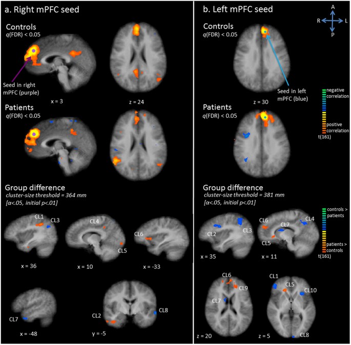


Exploration Of The Brain In Rest Resting State Functional Mri Abnormalities In Patients With Classic Galactosemia Scientific Reports


3 Dimensional Brain Mri Segmentation Based On Multi Layer Background Subtraction And Seed Region Growing Algorithm Scientific Net
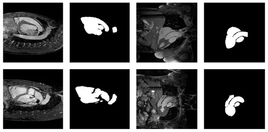
Researchers placed seeds in dark 5 mm NMR test tubes with either water or agar at °C to test germination of the rapeseed The investigators noted that 2% agar resulted in good seedtoexteriorFrom the seed region to capture the whole left atrium The experimental results demonstrate the accuracy and robustness of our approach I INTRODUCTION Automatic segmentation of the left atrium in magnetic resonance imaging (MRI) is a challenge and important task in medical image analysis For example, it can be used to analyze atrial The DLPFC a priori seed region (±36, 27, 29) was identified as less active in depressed participants during task performance in an emotioninterference, "conflict" matching task The a priori precuneus seed (±7, −60, 21) was chosen based on the literature (12, 38)


Post Mortem Mapping Of Connectional Anatomy For The Validation Of Diffusion Mri Biorxiv


Looking At Connections Between Brain Regions 1 Some


Resting State Functional Mri Everything That Nonexperts Have Always Wanted To Know American Journal Of Neuroradiology


Diffusion Tensor Imaging Guided Resection Radiology Key
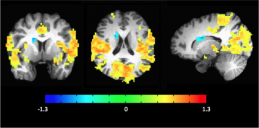


Mri Resting State Fmri Carney Institute For Brain Science Brown University
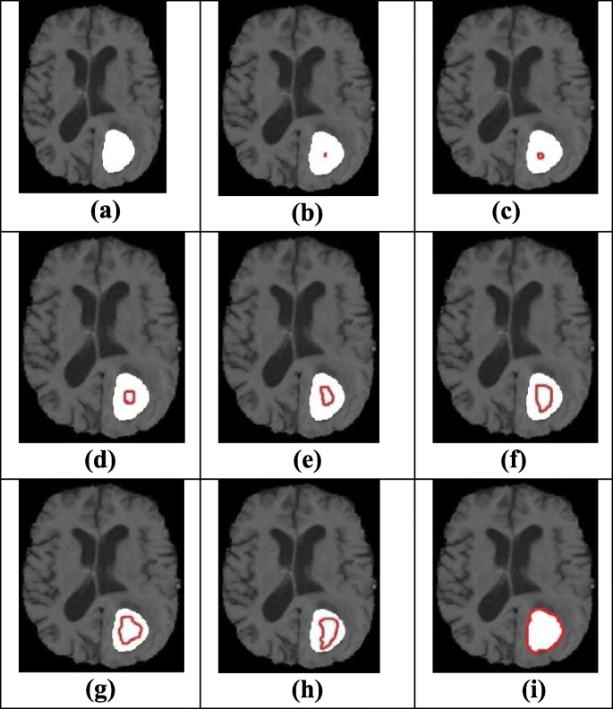


Morphological Edge Detection And Brain Tumor Segmentation In Magnetic Resonance Mr Images Based On Region Growing And Performance Evaluation Of Modified Fuzzy C Means Fcm Algorithm Springerlink


1


Network Analysis Of The Default Mode Network Using Functional Connectivity Mri In Temporal Lobe Epilepsy Protocol


Table Xi From Seed Based Region Growing Sbrg Vs Adaptive Network Based Inference System Anfis Vs Fuzzyc Means Fcm Brain Abnormalities Segmentation Semantic Scholar


Seed Regions Of The Seed Based Resting State Analysis Seed Regions And Download Scientific Diagram
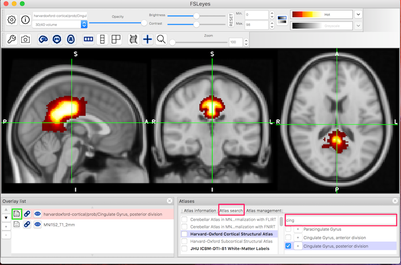


Fsl Fmri Resting State Seed Based Connectivity Neuroimaging Core 0 1 1 Documentation


Identifying Resting State Networks From Fmri Data Using Icas By Gili Karni Towards Data Science


Presurgical Resting State Functional Mri Language Mapping With Seed Selection Guided By Regional Homogeneity Hsu Magnetic Resonance In Medicine Wiley Online Library
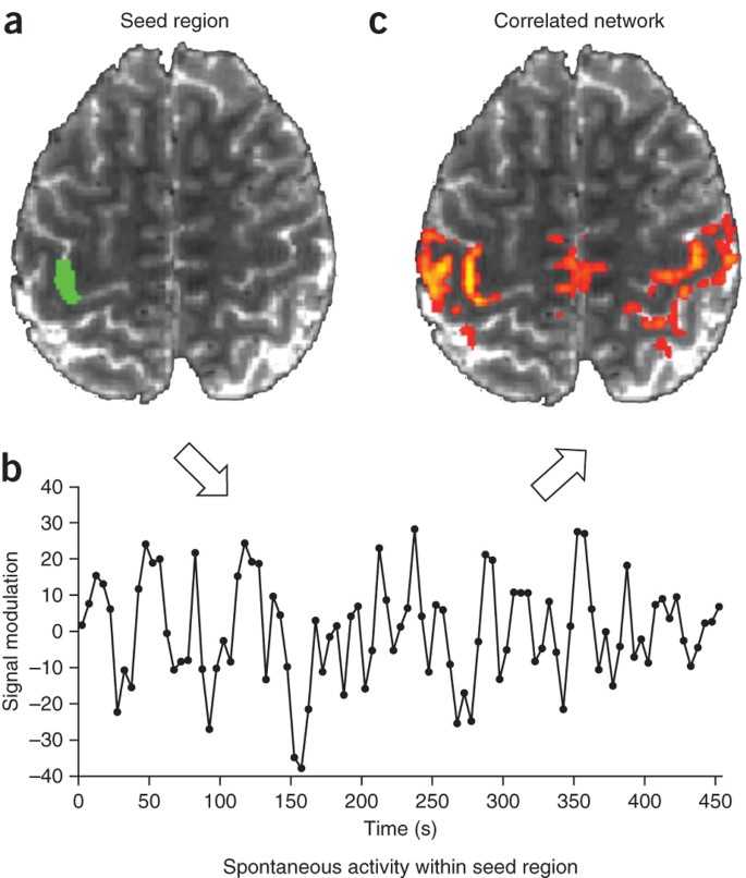


Opportunities And Limitations Of Intrinsic Functional Connectivity Mri Nature Neuroscience


Anatomical And Functional Organization Of The Human Substantia Nigra And Its Connections Biorxiv


Fmri


Mriquestions Com Uploads 3 4 5 7 Heuvel Reviewbrainnets1 Pdf
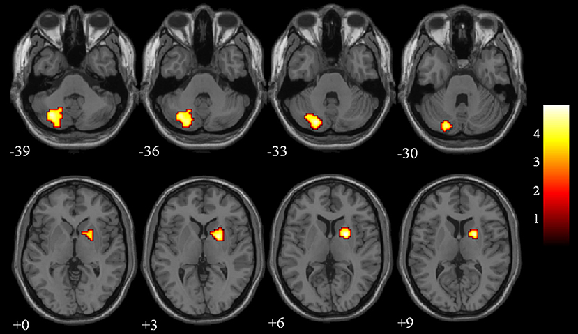


Frontiers Aberrant Brain Regional Homogeneity And Functional Connectivity Of Entorhinal Cortex In Vascular Mild Cognitive Impairment A Resting State Functional Mri Study Neurology


Brain Structure And Function In School Aged Children With Sluggish Cognitive Tempo Symptoms Journal Of The American Academy Of Child Adolescent Psychiatry


Resting State Fmri An Overview Sciencedirect Topics


How To Safely Perform Magnetic Resonance Imaging Guided Radioactive Seed Localizations In The Breast Journal Of Clinical Imaging Science


Weak Functional Connectivity In The Human Fetal Brain Prior To Preterm Birth Scientific Reports
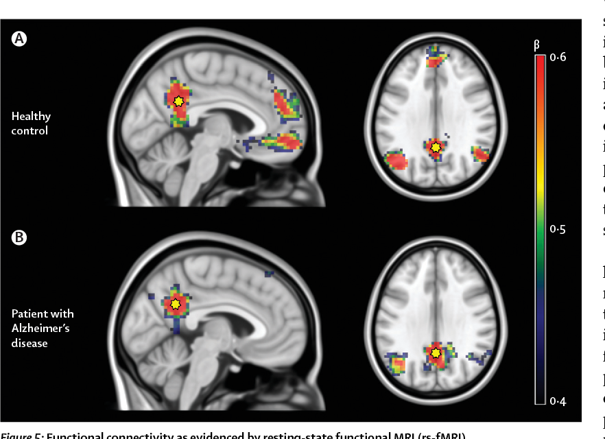


Figure 5 From Multimodal Imaging In Alzheimer S Disease Validity And Usefulness For Early Detection Semantic Scholar


Neuropsychiatric Neurologic Disorder Classification Prediction Brain Signal Processing Lab


A Seed And Target Region Of Interest Roi Of The Corticospinal Tract Download Scientific Diagram


Resting State Functional Mri Everything That Nonexperts Have Always Wanted To Know American Journal Of Neuroradiology


A Novel Magnetic Resonance Imaging Segmentation Technique For Determining Diffuse Intrinsic Pontine Glioma Tumor Volume In Journal Of Neurosurgery Pediatrics Volume 18 Issue 5 16


1


Diffusion Tensor Imaging Dti Fiber Tracking Imagilys


Correlation Of Copper Measurements With Functional Connectivity Mri Download Scientific Diagram


J Imaging Free Full Text Enhanced Region Growing For Brain Tumor Mr Image Segmentation


Diagnostics Free Full Text The Seven Deadly Sins Of Measuring Brain Structural Connectivity Using Diffusion Mri Streamlines Fibre Tracking Html


A Novel Mri Marker For Prostate Brachytherapy International Journal Of Radiation Oncology Biology Physics


Sleep Deprivation Increases Dorsal Nexus Connectivity To The Dorsolateral Prefrontal Cortex In Humans Pnas


Frontiers Differences In Cortical Structure And Functional Mri Connectivity In High Functioning Autism Neurology


Plos One Continuous Descending Modulation Of The Spinal Cord Revealed By Functional Mri


The Role Of Resting State Functional Mri For Clinical Preoperative Language Mapping Cancer Imaging Full Text


Defining The Seed Region Of Interest Ppi Analysis Investigates Download Scientific Diagram


Resting State Functional Mri In Depression Unmasks Increased Connectivity Between Networks Via The Dorsal Nexus Pnas


Frontiers Resting State Fmri Reveals Increased Subthalamic Nucleus And Sensorimotor Cortex Connectivity In Patients With Parkinson S Disease Under Medication Frontiers In Aging Neuroscience


Ultra High Field Mri Reveals Mood Related Circuit Disturbances In Depression A Comparison Between 3 Tesla And 7 Tesla Biorxiv


Characterizing The Spectrum Of Task Fmri Connectivity Approaches Ppt Download


Intrinsic Functional Connectivity As A Tool For Human Connectomics Theory Properties And Optimization Journal Of Neurophysiology


Pet And Mri Show Differences In Cerebral Asymmetry And Functional Connectivity Between Homo And Heterosexual Subjects Pnas


Resting State Functional Mri Everything That Nonexperts Have Always Wanted To Know American Journal Of Neuroradiology
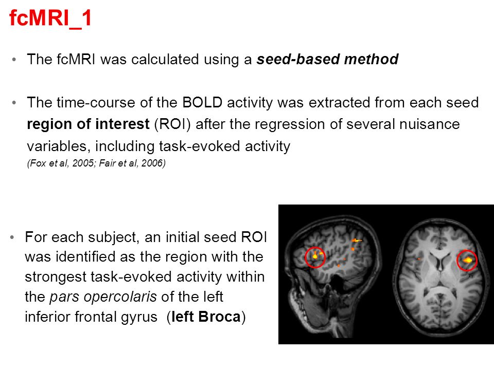


Functional Connectivity Mri In Patients With Brain Tumors Ppt Download


Mri Based Automated Detection Of Implanted Low Dose Rate Ldr Brachytherapy Seeds Using Quantitative Susceptibility Mapping Qsm And Unsupervised Machine Learning Ml Radiotherapy And Oncology


2


Mapping Cognitive And Emotional Networks In Neurosurgical Patients Using Resting State Functional Magnetic Resonance Imaging In Neurosurgical Focus Volume 48 Issue 2
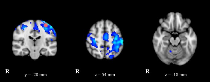


Cerebello Thalamo Cortical Network Is Intrinsically Altered In Essential Tremor Evidence From A Resting State Functional Mri Study Scientific Reports
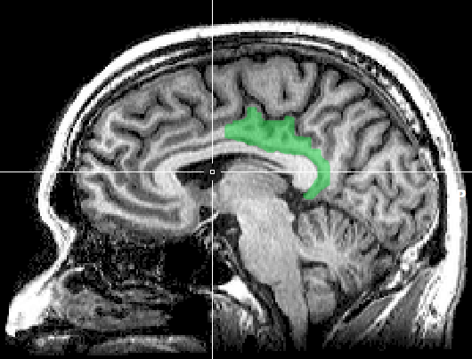


Posterior Cingulate Cortex Wikipedia
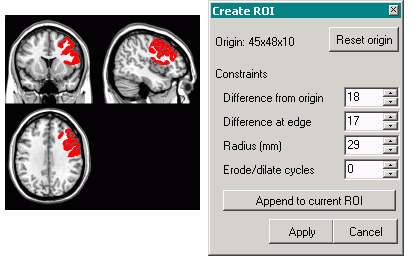


Creating 3d Rois With Mricro


The Top Image Shows A Slice From A Clinical Mri Scan Along With The Download Scientific Diagram


Development Of Functional Connectivity Voxelwise Resting State Download Scientific Diagram


Exploration Of The Brain In Rest Resting State Functional Mri Abnormalities In Patients With Classic Galactosemia Abstract Europe Pmc


The Role Of Resting State Functional Mri For Clinical Preoperative Language Mapping Cancer Imaging Full Text


Link Springer Com Content Pdf 10 1007 2f978 3 319 567 2 9077 1 Pdf


Resting State Fmri An Overview Sciencedirect Topics


Machine Learning In Resting State Fmri Analysis Sciencedirect


Magnetic Resonance Image Guided Brachytherapy Abstract Europe Pmc


Quantitative Dynamic Thoracic Mri Application To Thoracic Insufficiency Syndrome In Pediatric Patients Radiology


Love Related Changes In The Brain A Resting State Functional Magnetic Resonance Imaging Study Topic Of Research Paper In Clinical Medicine Download Scholarly Article Pdf And Read For Free On Cyberleninka Open Science


Resting State Functional Mri Everything That Nonexperts Have Always Wanted To Know American Journal Of Neuroradiology


Resting State Functional Mri Everything That Nonexperts Have Always Wanted To Know American Journal Of Neuroradiology


Breast Mri Tumour Segmentation Using Modified Automatic Seeded


Mathematics Free Full Text Feasibility Of Automatic Seed Generation Applied To Cardiac Mri Image Analysis Html


Ismrm Digital Posters Page Interventional Interventional Mr And Safety Issues


Breast Mri Tumour Segmentation Using Modified Automatic Seeded


Figure 1 From Texture Feature Based Automated Seeded Region Growing In Abdominal Mri Segmentation Semantic Scholar


Defining The Seed Region Of Interest Ppi Analysis Investigates Download Scientific Diagram


Resting State Functional Magnetic Resonance Imaging Connectivity Between Semantic And Phonological Regions Of Interest May Inform Language Targets In Aphasia Journal Of Speech Language And Hearing Research


Language Lateralization From Task Based And Resting State Functional Mri In Patients With Epilepsy Rolinski Human Brain Mapping Wiley Online Library


Concepts And Principles Of Clinical Functional Magnetic Resonance Imaging Chapter 13 The Cambridge Handbook Of Research Methods In Clinical Psychology


Level Set Based Hippocampus Segmentation In Mr Images With Improved Initialization Using Region Growing


Main Brain Areas Implicated In Resting State Functional Mri Diagram Of Download Scientific Diagram


Fsl Fmri Resting State Seed Based Connectivity Neuroimaging Core 0 1 1 Documentation


Philarchive Org Archive Lindoa 8


The Role Of Resting State Functional Mri For Clinical Preoperative Language Mapping Cancer Imaging Full Text


Performing Region Growing On An Mri Bone Image A Original Images Download Scientific Diagram


A Texture Based Tumor Detection And Automatic Segmentation


Altered Functional Connectivity In Post Ischemic Stroke Depression A Resting State Functional Magnetic Resonance Imaging Study European Journal Of Radiology


Resting State Fmri Wikipedia


0 件のコメント:
コメントを投稿